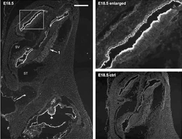
Figure 4.
ASIC2 immunolabeling results of a prenatal cochlear sections (E18.5). The negative control (E18.5 ctrl) was obtained by preincubating the primary antibody with antigenic peptide. The enlarged view given in the top right panel shows detailed labeling patterns in areas of the cochlea outlined by a frame. Arrow 1 points to the location of SG neurons, and the arrowhead indicates developing macula of utricle. Scale bar, ∼800 μm. SV, Scala vestibule; SM, scala media; ST, scala tympani.

