Figure 5.
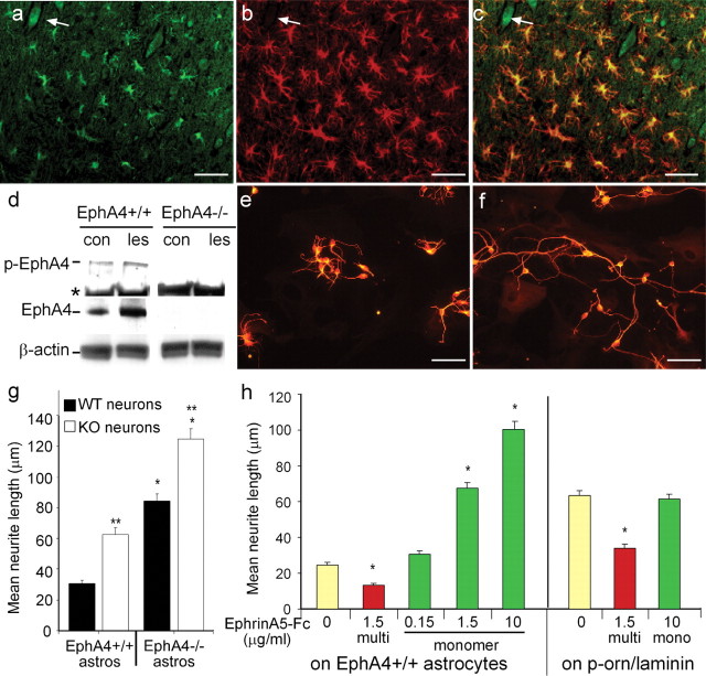
Expression of EphA4 on astrocytes inhibits neurite outgrowth. After spinal hemisection, EphA4 (a) and GFAP (b) are coexpressed as assessed by immunofluorescence on reactive astrocytes at the lesion site (c; a merged image of a and b). EphA4 was also expressed on some neurons (a-c, arrow). Western blot analysis (d) showed upregulation and phosphorylation of EphA4 (p-EphA4) at the lesion site (les) in comparison with unlesioned control (con) mice; * shows a nonspecific band present in all lanes. β-Actin was used as a loading control and EphA4-/- spinal cord as an EphA4 expression control. The EphA4 expression on astrocytes was inhibitory to cortical neuronal neurite outgrowth, because βIII-tubulin-positive cortical neurons on EphA4-/- astrocytes (e, g) had significantly longer neurites than on EphA4-expressing (EphA4+/+) astrocytes (f, g) after 22 hr (*p < 0.0001). EphA4-/- neurite outgrowth was also enhanced on EphA4-/- and EphA4+/+ astrocytes, compared with that of wild-type neurons (g; **p < 0.0001). h, The inhibition of neurite outgrowth by EphA4 on astrocytes could be blocked in a dose-dependent manner by addition of monomeric (mono) EphrinA5-Fc, but this had no effect on neurites grown on laminin. Multimerized (multi) EphrinA5-Fc inhibited neurite outgrowth both on astrocytes and on laminin (*p < 0.0001). Scale bars, 50 μm.

