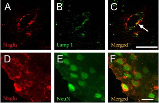
Figure 5.
NaGlu accumulated in neurons. Slice 6 from the injected hemisphere of mouse 23 was processed for cryosection (slice IL6, mouse 23 on Fig. 3). NaGlu was revealed by confocal fluorescence microscopy using a rabbit anti-human NaGlu serum (A, D). The anti-NaGlu (A) and lysosome-specific anti-Lamp1 (B) antibodies essentially stained different cytoplasmic organelles, although signals might occasionally colocalize (arrow in C). Cells showing cytoplasmic NaGlu signals (D) coexpressed the nuclear neuronal marker NeuN (E, F). Scale bar, 25 μm.

