
Figure 8.
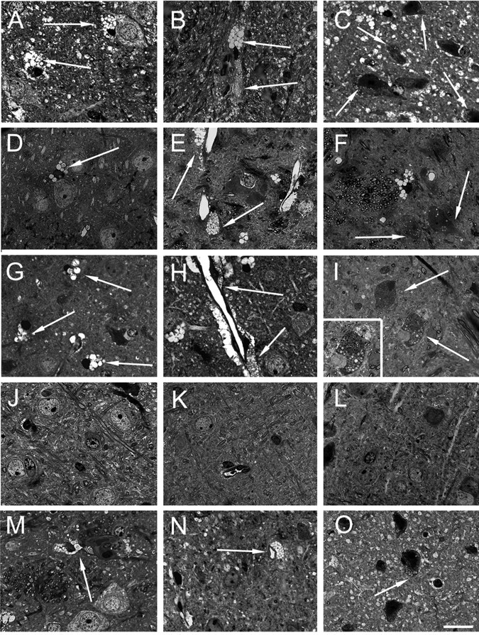
Disappearance or reduction of vacuolation in MPSIIIB mouse brain cells. Semithin sections stained with toluidine blue from brain slices of 6-week-old (A-C), 3-month-old (D-F), and 8-month-old (G-I) untreated MPSIIIB mice and from mouse 1 treated with AAV2-NaGlu vectors (J-L) and mouse 16 treated with AAV5-NaGlu vectors (M-O) are shown. Figure 4 indicates NaGlu activity in slices adjacent to those used for histopathology in these treated mice. Bright fine vacuoles in microglia (A) and larger and darker vacuoles in perivascular cells (B) were observed in the striatum of a 6-week-old untreated MPSIIIB mouse. Few neurons or astrocytes showed fine clear vacuoles in the parietal cortex (C). Large clear vacuoles were visible in microglia in the parietal cortex (D) and in microglia and perivascular cells in the putamen (E) of a 3-month-old untreated MPSIIIB mouse. Neurons or astrocytes with fine clear inclusions are visible in the putamen (F). Vacuoles in cortical microglia (G) and distension in perivascular cells near neurons or astrocytes with normal morphology (H) were observed in the putamen of an 8-month-old untreated mouse. Severely affected cells with abundant polymorphic vacuoles (I) as well as cells with picnotic nuclei (inset in I) were present at several other locations in the putamen of this mouse. Lysosomal distension was not observed in microglia (J), perivascular cells (K), or neurons or astrocytes (J-L) in the parietal cortex of the injected hemisphere of treated mouse 1 (slice 6 from the injected hemisphere). In contrast, residual lesions were observed in microglia (M) and perivascular cells (M, N) in the putamen of mouse 16 (slice 8 from the injected hemisphere). Vacuoles were also observed in neurons or astrocytes in the parietal cortex of the noninjected hemisphere (slice 8) of this mouse. Scale bar, 25 μm.