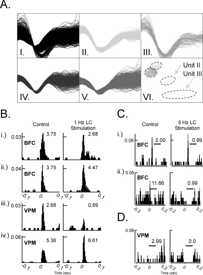
Figure 1.
Responses of individual neurons within the thalamus or cortex to whisker pad stimulation before and during LC stimulation. A, Off-line verification that unit waveforms originated from a single neuron. Recorded electrical activity (AI) from a single microwire electrode was discriminated as four unit waveforms (AII-AV) and represented in a scatter plot (PC1 vs PC2; AVI). Using our off-line criteria, Units II and III were verified as originating from a single neuron. B, The PSTHs illustrate spontaneous and somatosensory stimulus-evoked discharge of four simultaneously recorded neurons (1 per each row), before (left) and during (right) 1.0 Hz LC stimulation. Each histogram sums unit activity during an equal number of stimulus presentations. Bi and Bii are cortical neurons (BFC); Biii and Biv are thalamic neurons (VPM). During periods of LC activation, sensory-evoked excitation was suppressed (Bi, Biii) or facilitated (Bii, Biv) in cortical and thalamic neurons, respectively. Inset numbers represent the summed probability that the neuron will discharge in response to whisker pad stimulation. C, D, LC activation (5.0 Hz) also elicited an enhancement of postexcitatory- and stimulus-bound (Ci, Cii, D) inhibition in cortical (Ci, Cii) and thalamic (D) neurons, respectively. Inset numbers in C and D represent the summed probability that the neuron would discharge during the period indicated by the horizontal bar. In each histogram, the y-axis represents the probability that the neuron discharged for a given 1 msec bin (x-axis). Whisker pad stimuli were presented at 0.0 sec.

