
Figure 5.
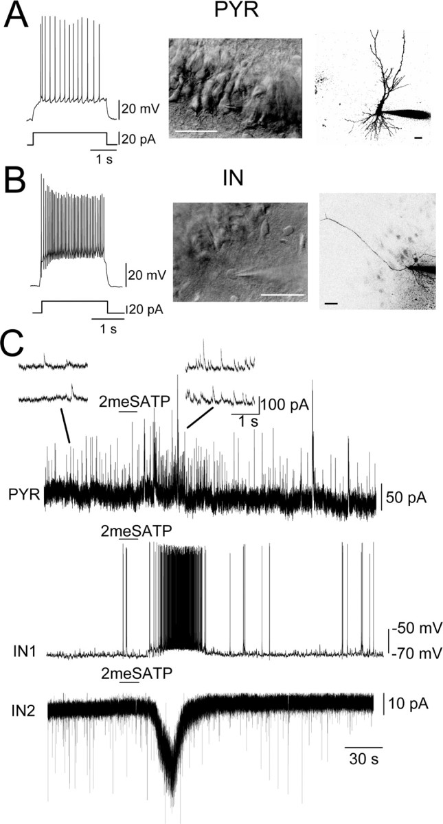
2meSATP excited interneurons in the stratum oriens in the CA3. A, An example of a pyramidal (PYR) neuron. Left trace, The membrane potential response to a 20 pA depolarizing current injection. Middle, An IR-DIC image of the CA3 region, from which the pyramidal neuron (PYR) showing the responses in A and C was recorded (the cell with the patch pipette tip). Scale bar, 50 μm. Right, A confocal image of the pyramidal neuron and the tip of a patch pipette filled with AlexaFluor 568. Scale bar, 50 μm. B, Left trace, Membrane potential response to a 40 pA depolarizing current injection. Middle, An IR-DIC image showing the interneuron (IN) recorded in the stratum oriens (the cell with the patch pipette tip). Scale bar, 50 μm. Right, A confocal image of the interneuron and the tip of a patch pipette filled with AlexaFluor 568. Scale bar, 50 μm. C, PYR, Membrane currents of the CA3 pyramidal neuron recorded with low-Cl internal solution at a holding potential of 0 mV. 2meSATP (100 μm) was applied at the horizontal bar. Consecutive time-extended versions of the membrane current are shown as insets above. IN1, The membrane potential of the interneuron shown in B. IN2, The membrane current of the interneuron in the stratum oriens. The holding potential was -70 mV. Note that the increase in IPSC frequency (PYR), the action potential generation (IN1), and the inward current (IN2) in response to 2meSATP were of highly similar time course.