
Figure 2.
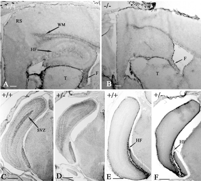
The posterior pole of the telencephalon. A, P7 WT mice. B, P7 -/- mice, parasagittal sections immunostained with GFAP. Cortical areas at the posterior pole are reduced in size in the p73-/- mouse. The white matter (WM) area is also smaller, and the hippocampal fissure (HF) is missing. F, Fimbria; RS, retrosplenial cortex; T, thalamus. C-F, The posterior pole of p73 heterozygous (D, F) and WT (C, E) mice at E18 in coronal Nissl (C, D) and Reelin (E, F) stained sections. Sections are at the same level to show the size reduction of the caudal telencephalon and the dorsal shift of entorhinal cortex. The proliferative subventricular zone (SVZ) is still visible in C (WT) but not in D (p73+/-). E, F, The number and distribution of Reelin-IR CR cells in the hippocampal fissure and in the cortical marginal zone are similar in WT and p73+/- mice. Scale bars, 200 μm.