
Figure 7.
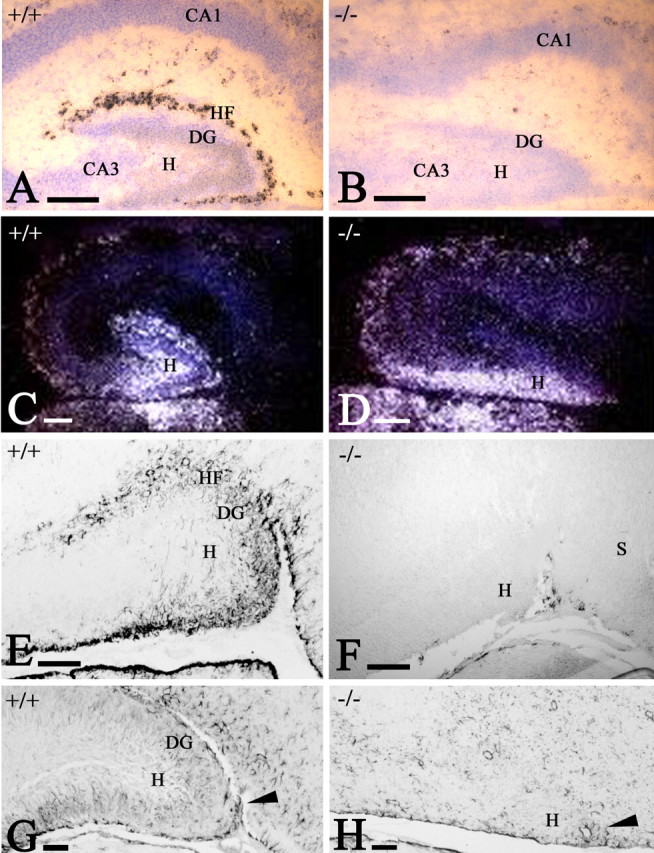
The postnatal hippocampal fissure (HF). A, B, Reelin ISH in WT (A) and p73-/- (B) mice at P2. B, CR cells are absent, and the molecular layers of dentate gyrus (DG) and CA1/CA2 are fused. C, D, Calretinin ISH in WT (C) and p73-/- (D) mice at P2. In the mutant, calretinin-positive hilus (H) cells have abnormal distribution. E, F, GFAP IHC in WT (E) and p73-/- (F) mice at P2. GFAP stains subpial astrocytes in the HF and the incipient radial glia scaffold in the dentate gyrus. In the mutant, astrocytes are found only along the medial brain surface. G, H, GFAP in WT (G) and p73-/- (H) mice at P12. The radial glia scaffold is fully established in the WT dentate; in the mutant, astrocytes are sparse and lack radial orientation. Arrowheads mark the entrance of the fissure, which fails to fold in the mutant. S, Subiculum. Scale bars: A, B, 200 μm; C-H, 100 μm.