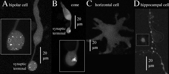
Figure 4.
Selective peptide-labeling of structures in the synaptic terminal of ribbon-containing neurons. Each picture shows planar projections of confocal optical sections through the entire depth of the cell. A, Bipolar cell. B, Cone photoreceptor. C, Horizontal cell. D, Cultured hippocampal neuron. The cell body of the labeled hippocampal neuron was above the field of view, and a labeled process followed the principal process of a nearby unlabeled cell, the position of which is indicated by the outline.

