Figure 5.

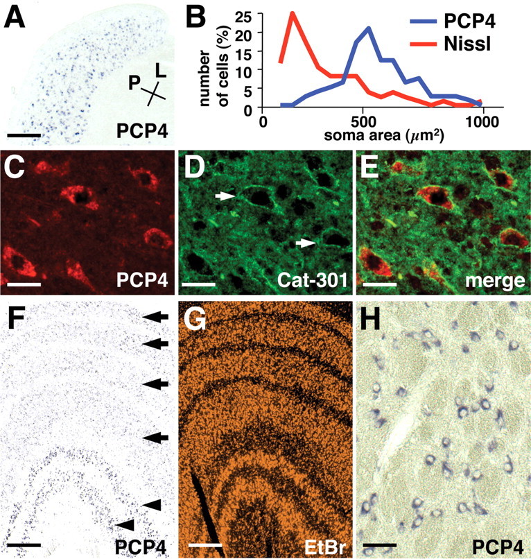
Expression of PCP4 in Y cells in the LGN. A, Expression of PCP4 mRNA in a horizontal section of the adult ferret LGN. PCP4-positive cells are abundant in A layers and less frequent in C layers. Conventions are as in Figure 3. Scale bar, 400 μm. B, The soma area of PCP4-positive cells (blue) and cells stained for Nissl substance (red) in the adult ferret LGN. C-E, Colocalization of PCP4 mRNA and Cat-301 immunoreactivity in the adult ferret LGN. LGN sections were double stained with in situ hybridization using a PCP4 probe (C) and immunohistochemistry using Cat-301 antibody (D). As reported previously, Cat-301 antibody recognizes perineuronal nets (arrows) of Y cells (D). PCP4-positive cells were surrounded by Cat-301-immunoreactive perineuronal nets (E). Scale bars: C-E, 30 μm. F, Expression of PCP4 mRNA in adult (2-3 years of age) macaque LGN revealed by in situ hybridization. PCP4 was strongly expressed in the magnocellular layers (arrowheads) but only weakly in the parvocellular layers (arrows). G, Ethidium bromide staining of the macaque LGN showing six layered structures. Scale bars: F, G, 400 μm. H, High-magnification view of PCP4-positive cells in the magnocellular layers in the macaque LGN. Scale bar, 50 μm.
