
Figure 8.
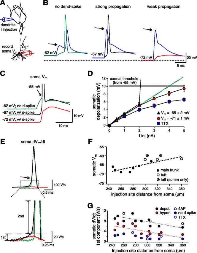
The somatic impact of dendritically initiated spikes. A, Experimental configuration. A dendritic electrode (blue traces) located 285 μm from the soma was used to inject current and record voltage, while the response at the soma was recorded with a second electrode. B, Effect of the Vm on signal propagation to the soma. Left, The injection of relatively small amounts of EPSC-shaped current (2 nA) does not evoke a dendritic spike but travels to the soma, where it provides enough slow depolarization to initiate a somatic action potential at this more depolarized Vm (-62 mV; green trace from the soma). The spike recorded in the dendritic trace is back-propagating. Middle, Larger current injections (4 nA) lead to the initiation of a dendritic spike and the subsequent generation of a short-latency somatic action potential at the normal Vm (-67 mV; black trace from the soma). Right, At hyperpolarized potentials (-72 mV; red trace from the soma), a dendritic spike is initiated by the large current injection (4 nA), but it is unable to propagate to the soma effectively enough to evoke an action potential output. C, Superimposition of the somatic recordings from B. Notice the apparently lower threshold for the spike evoked at -67 mV and the fast iEPSP rise time with the Vm at -72 mV. D, I-V plot of the depolarization recorded at the soma after dendritic current injections at the normal Vm (black triangles; n = 5), at hyperpolarized potentials (red circles; n = 7), and in the presence of TTX (blue squares; n = 4). The gray line shows the action potential threshold for slow somatic depolarizations without dendritic spikes with respect to the average Vm (-65 ± 1 mV; n = 5). E, First derivative (dV/dt) of the traces in C. The enlargement (bottom) shows the initial ramp component that is only present when a dendritic spike is evoked (black and red traces). The plot shows that this initial ramp can be further broken into two separate components: one that is larger and present only when propagation is strong enough to evoke action potential output (labeled second; black trace; shaded region), and another that is present regardless of a somatic spike (labeled first; black and red traces). F, Plot of the most hyperpolarized Vm at which a dendritic spike could still evoke a short-latency somatic output (<2 msec). Short-latency action potential output could not be evoked in two neurons when dendritic current injection initiated a spike in the apical tuft region. In these cells, action potential threshold at the soma was reached at the peak of the iEPSP (∼7 msec) and was therefore considered to be the result of summation (summ only) and not the direct result of the strong propagation of the dendritic spike. G, The amplitude of the first component of the initial dV/dt ramp depends on the distance of the injection site from the soma and other factors influencing spike propagation. The second component of the initial dV/dt ramp exhibited a similar location dependence (data not shown).