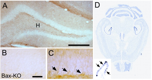
Figure 2.
Bax-IR in the DG of 2-month-old WT mice (A). Note that weak but significant Bax-IR was observed in a subset of cells localized in the SGZ (C, arrows). H indicates the hilar region of the hippocampal formation. Bax-KO DG did not show significant Bax-IR (B). DG from three independent animals were observed, and a representative image is shown. D, Insitu hybridization of Bax mRNA in the adult (2-month-old) WT mouse. Enlarged image of the DG shows a subset of cells that express higher amounts of Bax mRNA. Scale bars: A, 500 μm; B, C, 50 μm.

