
Figure 7.
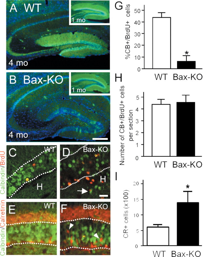
Reduction of CB immunoreactivity (green) in 4-month-old Bax-KO mice (A, WT versus B, Bax-KO). Insets in A and B demonstrate an apparent normal pattern of CB-IR in 1-month-old WT and Bax-KO mice. Nuclei were visualized by Hoechst33342 (blue). Scale bar, 200 μm. C, D, One month after BrdU injection, BrdU+ cells (red) double labeled with CB (green) were visualized. Note that most BrdU+ cells in the Bax-KO mice were CB-. Dotted lines indicate the boundary of the GCL. Scale bar, 50 μm. Arrow in D indicates CB+ neurons localized in the hilus. E, F, Double labeling for CB (green) and CR (red) in 4-month-old WT (E) and Bax-KO (F) mice. In the Bax-KO, there were many CR+ cells in the middle of the GCL (arrowhead). G, Quantification of the percentage of double-labeled cells in the two groups. More than 200 BrdU-labeled cells from a similar region (caudal hippocampal) were examined per mouse. *p < 0.05 in t test comparison of WT versus Bax-KO mice. H, The number of double-labeled cells per section in the DG. I, The total number of CR+ cells in every sixth section through the DG (n = 4).