
Figure 2.
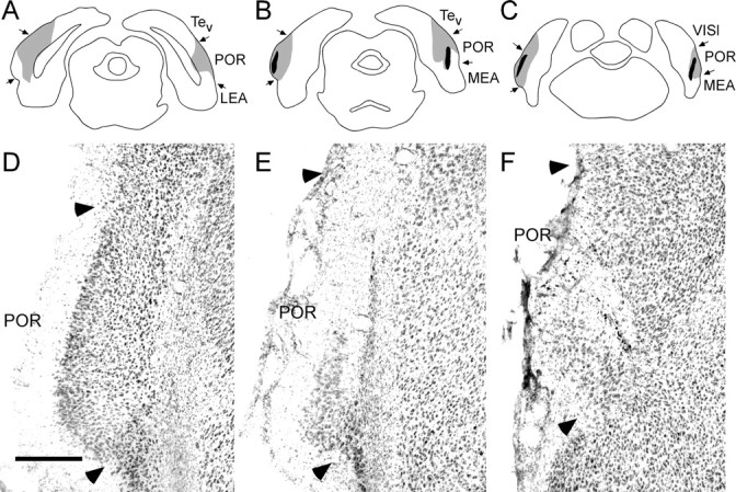
Postrhinal lesions. A-C, Schematic of the placement of the largest (gray) and smallest (black) POR neurotoxic lesion at three rostrocaudal levels, -7.6, -8.2, and -8.7 mm, according to bregma (Paxinos and Watson, 1998). D-F, Photomicrograph of a representative POR neurotoxic lesion. Arrowheads indicate cytoarchitectonic boundaries of POR. LEA, Lateral entorhinal cortex; MEA, medial entorhinal cortex; Tev, temporal cortex; VISl, lateral visual association cortex. Scale bar, 300 μm.