
Figure 7.
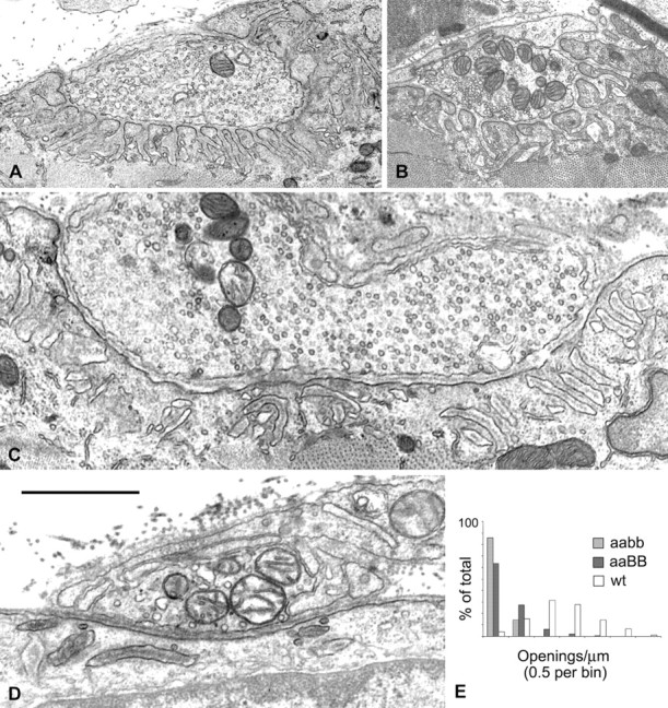
Electron microscopy of junctional folds. A, Nerve-muscle contacts in a β2-syntrophin null mouse and its wild-type littermate (B) show numerous, well organized junctional folds and associated openings to the synaptic cleft. C, D, Nerve-muscle contacts from α/β2-syntrophin null mice. C, A nerve-muscle contact of mature appearance. Approximately 20 fold-like structures are shown. Many are disorganized (left two-thirds of the image), but even where they are reasonably organized (right one-third), there are no openings to the synaptic cleft in the section shown. D, A contact of immature appearance. Disorganized fold structures are apparent just below the muscle cell surface. The central dark stripes within the fold structures in C and D are basal lamina. Scale bar: A, 2.5 μm; B, 1.8 μm; C, 1.2 μm; D, 1.0 μm. E, The values for openings to the synaptic cleft per micrometer of presynaptic membrane for all nerve-muscle contacts are grouped by bins of 0.5 for two α/β2-syntrophin null mice (this study) and α-syntrophin null mice and their wild-type littermates (Adams et al., 2000).