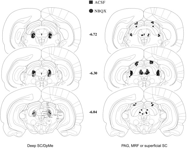
Figure 1.
The locations of the tips of the infusion cannulas in the deep SC/DpMe, the dorsal/lateral PAG, the lateral MRF, or the superficial SC. The closed squares represent the cannula placements with ACSF infusion, and the closed circles represent the cannula placements with NBQX infusion. In the deep SC/DpMe, because of a large number of the cannulas implanted, the cannula placements are generally illustrated with the shaded area (left) with some squares and circles that are an example of the infusion of ACSF and 0.79 nmol of NBQX. The right panel illustrates the cannula placements in the PAG, the MRF, or the superficial SC, in which NBQX infusion was ineffective in blocking potentiated startle. MGM, Medial geniculate nucleus, medial part; SG, suprageniculate thalamic nucleus; DLPAG, the dorsolateral periaqueductal gray; DpG, deep gray layer of the superior colliculus; DpWh, deep white layer of the superior colliculus; IMLF, interstitial nucleus of the medial longitudinal fasciculus; InG, intermediate gray layer of the superior colliculus; InWh, intermediate white layer of the superior colliculus; LPAG, lateral periaqueductal gray; MGD, medial geniculate nucleus, dorsal part; MGV, medial geniculate nucleus, ventral part; Op, optic nerve layer of the superior colliculus; PIN, posterior intralaminar thalamic nucleus; PaR, pararubral nucleus; SuG, superficial gray layer of the superior colliculus; VPAG, ventral periaqueductal gray.

