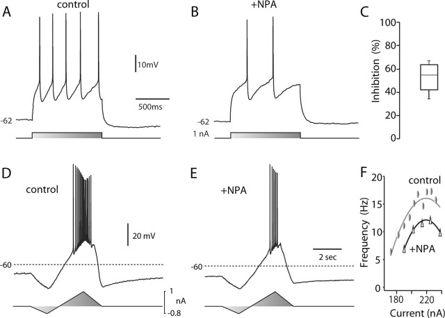
Figure 1.
D2 receptor activation reduced evoked activity. A, Repetitive spiking evoked in a cholinergic interneuron by intrasomatic current injection during a recording in a tissue slice. B, The same neuron after bath application of NPA (10 μm). Note the reduction in evoked discharge. C, Statistical summary of NPA-induced reduction in evoked spiking. D, Repetitive activity evoked by somatic injection of a current ramp. E, The response to the same stimulus was reduced after application of NPA (10 μm). F, Instantaneous discharge frequency-current injection plot for the neuron shown in D and E. Data were fit with a polynomial function: control, -206.8 + 2.03i -0.0046i2; NPA, -287.4 + 2.74i -0.0062i2, where i is current. Similar results were obtained in two other neurons tested. Triangles indicate NPA application.

