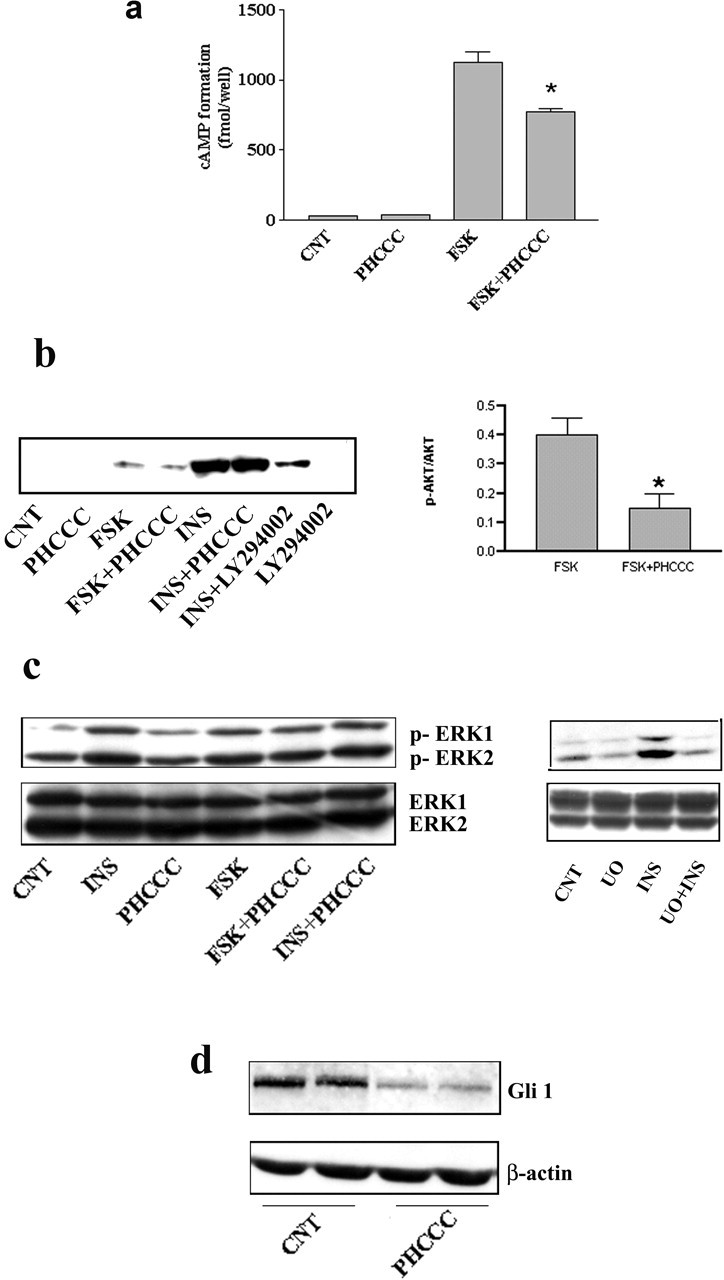
Figure 7.

a, PHCCC reduces forskolin-stimulated cAMP formation in cultured granule cells. The Figure refers to data obtained in cultures incubated in Krebs-Henseleit buffer. Similar results were obtained in cultures incubated in their growing medium (BME alone) both at 4 and 24 hr after plating (data not shown). Under no conditions was PHCCC able to reduce basal cAMP formation. CNT, Control; PHCCC, 30 μm; forskolin, 10 μm; values are means ± SEM of six determinations. *p < 0.05 (1-way ANOVA plus Fisher's PLSD) versus forskolin alone. b, Western blot analysis of phosphorylated AKT in cultures plated in BME alone and treated for 10 min with the indicated drugs. CNT, Control; forskolin (10 μm); insulin (0.5 mg/ml); LY294002 (10 μm) was applied 30 min before insulin. Densitometric analysis of values obtained with forskolin alone and forskolin plus PHCCC (n = 4) are also shown. Values (normalized by the expression of nonphosphorylated AKT) are means ± SEM. *p < 0.05 (Student's t test) versus forskolin alone. Representative immunoblots of phosphorylated and nonphosphorylated ERK1/2 in cultures challenged with the indicated drugs are shown in c. Incubation times and concentrations are the same as in b. UO, UO126 (10 μm). Note that PHCCC was unable to affect the MAPK pathway and that UO126 slightly reduced the basal levels of phosphorylated ERK1/2 and more substantially reduced the activation of the MAPK pathway by insulin. Blots were repeated three times with similar results. Expression of the transcription factor Gli-1 in granule cells treated with PHCCC (30 μm) for 30 min is shown in d. CNT, Control. A reduced expression of Gli-1 was consistently seen in four additional samples per group.
