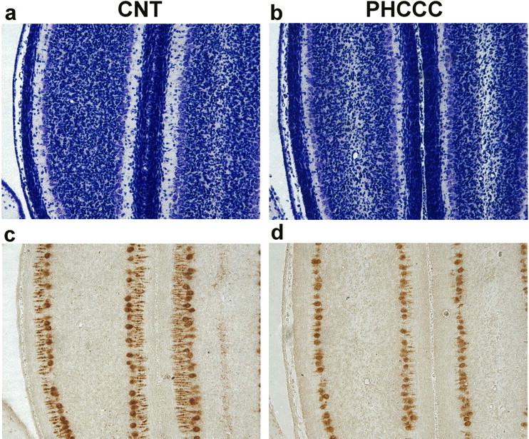
Figure 8.

Nissl staining (a, b) and calbindin immunostaining (c, d) of comparable cerebellar sections from rats treated intracerebrally with vehicle or with PHCCC (5 nmol/2 μl/2 min infused into the cerebellar region every other day from P3 to P9). Animals were killed at P10. Note the cell reduction in the internal granular layer in the representative cerebellar section of a PHCCC-treated animal (b). The same animal shows Purkinje cells with a dystrophic dendritic tree, as revealed by the calbindin immunostaining in d. These morphological abnormalities were observed in all PHCCC-treated animals.
