
Figure 1.
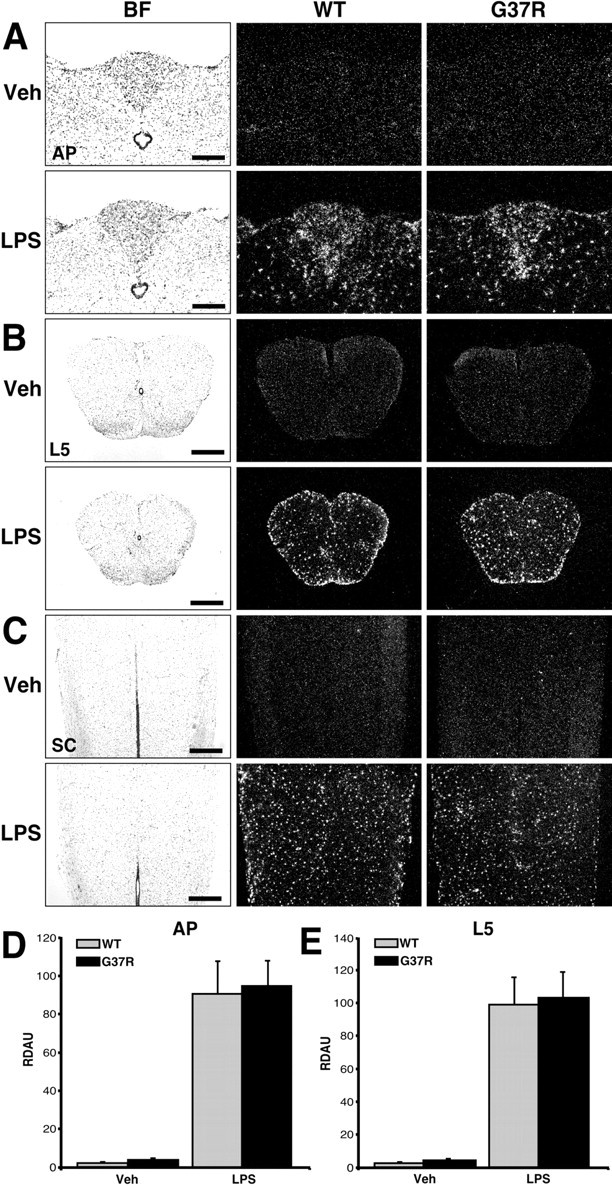
Effect of a single bolus of LPS on TLR2 gene expression in the brains and spinal cords of SOD1G37R mice and their WT littermates. These bright-field (BF) and dark-field photomicrographs depict the expression pattern of TLR2 mRNA 24 hr after a single intraperitoneal injection of vehicle (Veh) or LPS (1 mg/kg of body weight). Coronal and longitudinal sections were hybridized using a mouse TLR2 cRNA probe and dipped into NTB2 emulsion milk. A, Coronal sections at the level of the AP. B, Coronal sections within the L5 segment of the spinal cord (SC). C, Longitudinal slices of the SC. Note the strong and similar hybridization signal within the brain and spinal cord of both WT and SOD1G37R mice that received an intraperitoneal bolus of LPS. Semiquantitative analysis was performed in regions of the AP (D) and L5 segment (E). Data are means ± SEM. The expression levels were comparable in the CNS of both mouse strains after the acute endotoxemia. Statistical analysis was performed by a two-way ANOVA, which indicated a significant main effect (p < 0.0001) between the vehicle- and LPS-treated groups. Scale bars: A, 200 μm; B, C, 500 μm. RDAU, Refraction density in arbitrary units. (Means ± SEM).