
Figure 4.
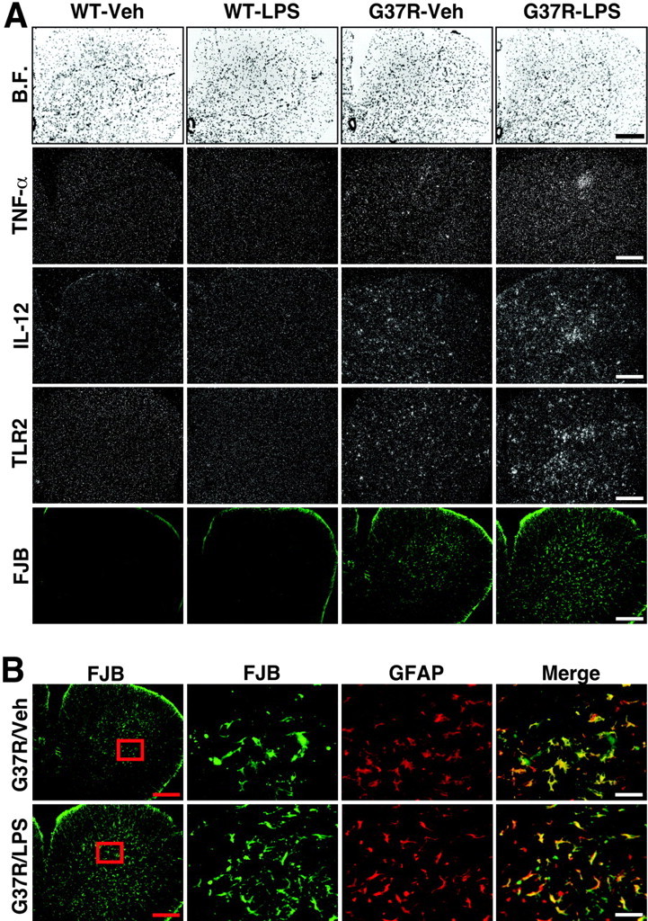
Robust inflammatory response in ventral spinal horn of chronically LPS-treated SOD1G37R mice associated with massive degeneration of astrocytes. The bright-field (B.F.) and dark-field photomicrographs depict representative examples of the hybridization signal for TNF-α, IL-12, and TLR2 mRNA in the L5 segment of the spinal cord (A). Here also the hybridization signal for IL-12 and TLR2 overlaps with the fluorochrome FJB. Neuronal cell bodies and axons contained FJB staining in this region. Please also note the robust FJB signal over astrocytes, a phenomenon that was specific to the L5 region (B). These data suggest an intimate link between degeneration of neurons and astrocytes in this region of spinal cord from SOD1G37R mice. Age of mice at time of analysis, 45-46 weeks. Scale bars: A and B (left panels), 500μm; B (high magnifications, merge), 50μm. See the legend to Figure 2 for definitions of abbreviations.