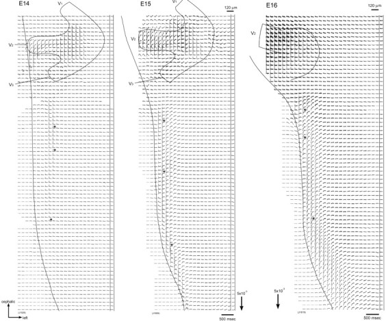
Figure 1.

Multiple-site optical recordings of trigeminal responses. Multiple-site optical recordings of neural responses were made in intact preparations of E14-E16 embryonic rat brainstems. The optical signals were evoked by applying a brief positive square current pulse (8 μA/5 msec) to the right maxillary (V2) nerve with a microsuction electrode. The evoked optical signals were detected from the ventral side of the brainstem in a single sweep. The 1020-site simultaneous recordings were made in four different contiguous regions by sliding the photodiode array over the image of the preparation. The direction of the arrows at the bottom indicates an increase in transmitted light intensity (decrease in dye absorption), and the length of the arrows represents the stated value of the fractional change ΔI/I. The signals indicated by asterisks are enlarged in Figure 2.
