Figure 2.
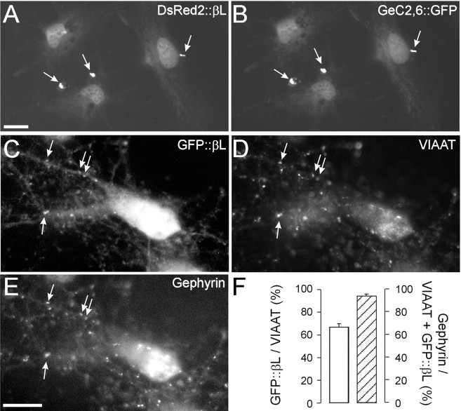
Accumulation of transfected GFP::βL opposite inhibitory synaptic terminals in DIV12 SCNs. A, B, COS-7 cells transfected with DsRed2::βL (A) and GeC2,6::GFP (B). DsRed2::βL is trapped by intracellular gephyrin aggregates and therefore can be regarded as capable of binding to gephyrin. C, Image of a neuron containing GFP::βL. D, E, Corresponding endogenous VIAAT (D) and gephyrin (E) immunoreactivity. F, Fraction of VIAAT-immunoreactive terminals that also contained GFP::βL (left y-axis) and fraction of VIAAT- and GFP::βL-immunoreactive synapses that also contained gephyrin (right y-axis). Note the high degree of colocalization between GFP::βL, VIAAT, and gephyrin. Scale bars, 10 μm.

