
Figure 3.
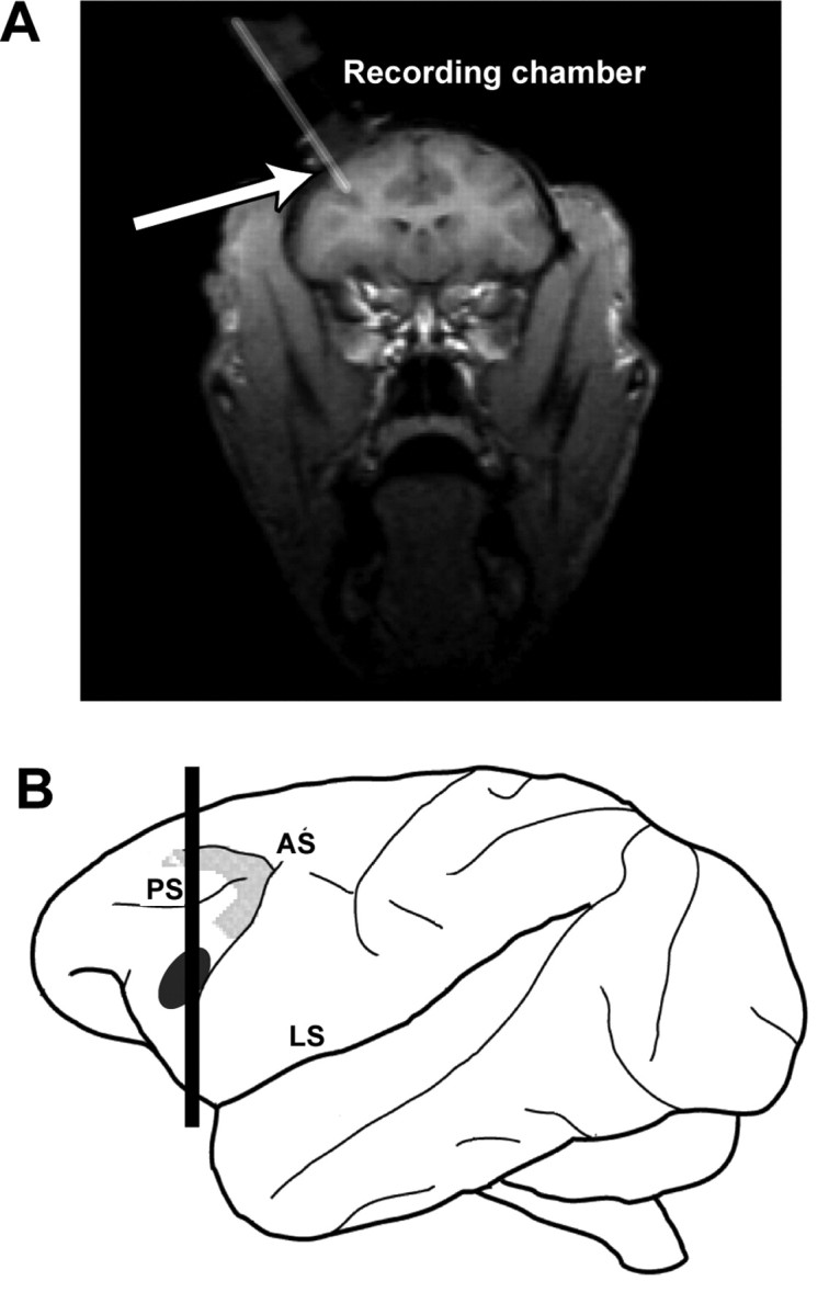
A, A coronal magnetic resonance image illustrating the approach of a recording electrode, as indicated by the arrow, through the recording chamber and into the vPFC. The approximate plane of section of this image is illustrated by the vertical black line through the schematic of the rhesus brain (B). The black ellipse on this schematic encompasses the region where we recorded auditory neurons from two rhesus monkeys, and the enclosed gray area outlines the approximate location of area 8a, a cortical area involved in auditory spatial processing (Russo and Bruce, 1994). AS, Arcuate sulcus; LS, lateral sulcus; PS, principal sulcus.