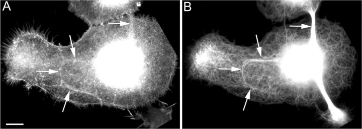
Figure 1.
Microtubules align with actin filament bundles during early axogenesis. Shown are images of neurons fixed and stained for fluorescence visualization of actin filaments (A) and microtubules (B) during early axonal outgrowth. Large lamellae developed in which microtubules and actin bundles formed along the axes of presumed future axons. Microtubules and actin bundles were seen to colocalize in some cases (see arrows in A and B). Scale bar, 10 μm.

