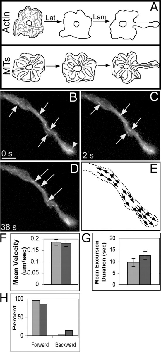
Figure 8.

Directionality of GFP-EB3 excursions in axons grown in the absence of actin filaments indicates uniform or nearly uniform microtubule polarity orientation. Neurons were cultured overnight in the presence of latrunculin to inhibit the formation of a filamentous actin network and then induced to grow axons the following day in the presence of the drug (for treatment regimen, see A). Axons extended in the absence of actin filaments were approximately half as long as control axons and were thicker in diameter. More GFP-EB3 “comets” were seen across the axon width (arrows in B-D). See movie 3 (available at www.jneurosci.org as supplemental material). The growth cone was essentially absent in the drug-treated axon, and instead appeared as a club-like region, dense in microtubule ends, at the distal end of the growing axon (arrowhead region denoted in B). The mean velocity was unchanged compared with control axons, whereas the duration of GFP-EB3 excursions increased (F, G; darker bars are actin-depleted data, and lighter bars are control data). The percentage of forward excursions was only slightly lower than in control axons, indicating that microtubules in the axon achieved uniform (or nearly uniform) plus-end distal polarity orientation in the absence of actin filaments (H). Scale bar, 5 μm. Lam, Laminin; Lat, latrunculin; MT, microtubule.
