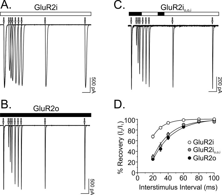
Figure 3.
Recovery from desensitization kinetics are sensitive to the exchange of three residues in GluR2i. A-C, Recovery from desensitization was evaluated by delivering pairs of 10 msec pulses of 1 mm glutamate separated by interstimulus intervals ranging from 20 to 500 msec. Traces represent superimposed responses from cells expressing GluR2i, GluR2o, and mutant GluR2i with exchanges of region 1 and 2 (GluR2io,o,i). Arrows denote on set of glutamate pulses. Calibration bar, 50 msec. D, The time course of recovery from desensitization was determined for each cell by measuring the amplitude of the second response (I2) relative to the first response (I1) of a pair of pulses and plotting these values as a function of interstimulus interval. The τREC for each cell was determined by fitting these recovery plots with a single exponential function. Statistically significant differences were found between τREC for GluR2i, GluR2o, and GluR2io,o,i (F(2,27) = 7.2; p < 0.005). Post hoc comparisons showed that although the τREC for GluR2io,o,i was not different from the τREC for GluR2o (p > 0.05), both differed from the τREC for GluR2i (p < 0.05).

