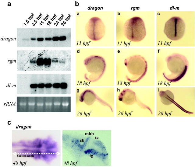
Figure 6.
Developmental expression of zebrafish dragon, rgm, and dl-m. a, Northern blot analysis showing zebrafish dragon, rgm, and dl-m mRNAs at different developmental stages as indicated (1.5-36 hpf). The bottom panel shows the corresponding 18S rRNA bands. b, In situ hybridization patterns of zebrafish dragon (a, d, g), rgm (b, e, h), and dl-m (c, f, i) mRNAs at the indicated developmental stages. c, A dorsal view and a longitudinal section of a 48-hr-old zebrafish embryo after in situ hybridization with a dragon probe. Significant staining is observed in the tectum (tc) and the tegmentum (tg) of the midbrain, the cerebellum (cb), and the hindbrain. mhb, Midbrain hindbrain boundary; hpf, hours postfertilization, 28.5°C.

