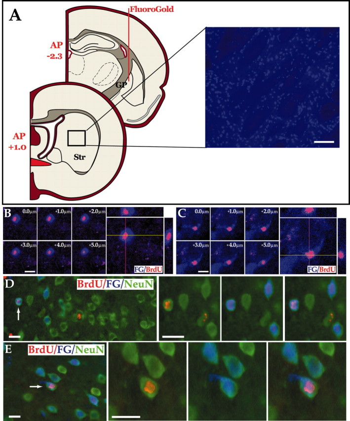
Figure 5.

Newly generated striatal neurons project to the globus pallidus. A, FG was injected into the ventral portion of the globus pallidus of rats that had received AdBDNF 6 weeks earlier. The rats were then killed 1 week later, and FG uptake by striatopallidal projection neurons was assessed. Diffusion of the retrograde tracer averaged 0.7 mm from the injection site at P -2.3, so that the most rostral extent of diffusion was typically to P -1.6 mm; no evidence of any diffusion of FG to the striatum was ever noted. Striatal sections were, thus, scored rostrally from AP +1.0, to avoid any possible diffusion artifact. Scale bar, 100 μm. B, C, Confocal images of FG+ (blue)/BrdU+ (red) double-immunolabeled cells in the striata of AdBDNF-injected rats (7 week survival), shown as both single optical sections and orthogonal views in the xz and yz planes, to confirm that scored BrdU+ cells were FG+. D, E, Two examples of BrdU+ (pink)/NeuN+ (green) striatal neurons identified in an AdBDNF-treated rat, 7 weeks after viral injection. These cells (arrows) incorporated FG (blue) injected into the globus pallidus. The triple-labeled cells represent newly generated striatal neurons that have extended fibers to the pallidal targets. Scale bar, 16 μm.
