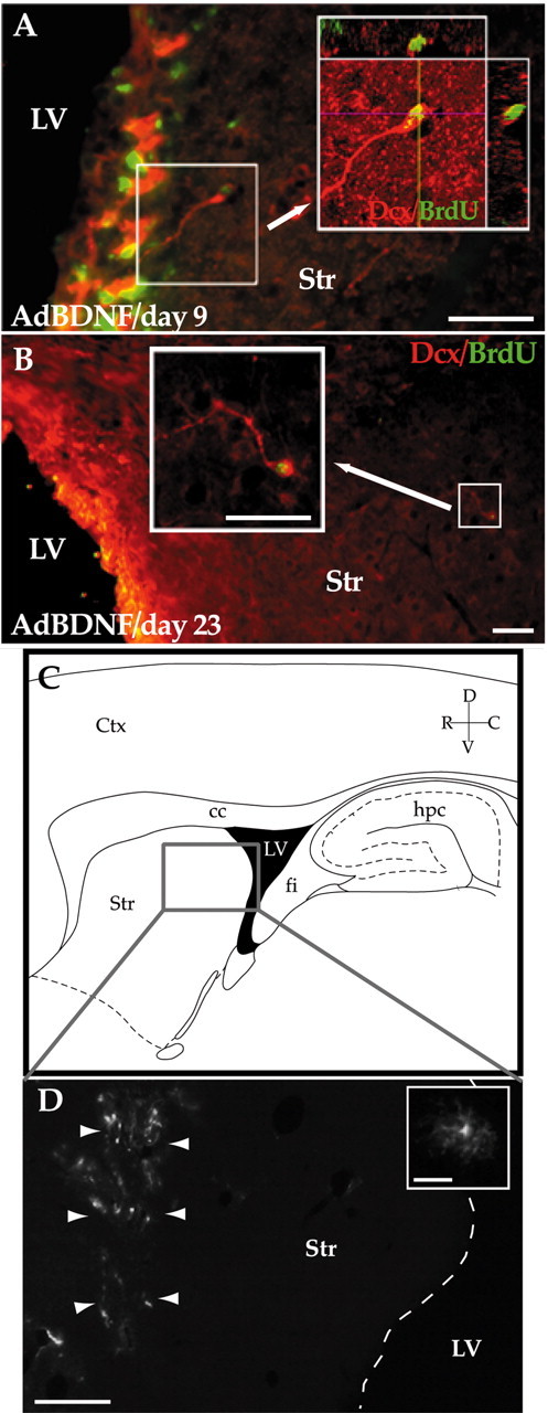
Figure 6.

AdBDNF-induced neurons arose from the SZ. A shows the neostriatal wall of a rat killed 9 d after AdBDNF that had been treated daily with BrdU for the week before sacrifice. Many Dcx+ (red)/BrdU+ (green) cells, likely neuronal migrants, were noted arising from the ventricular wall. No BrdU+/Dcx+ cells were seen beyond 100 μm from the ventricular wall at this time point. B, Low-power image of the striatum of a rat killed 23 d after AdBDNF that had been given BrdU for only the first week after viral injection. BrdU-tagged Dcx+ cells (arrow) were identified throughout much of the neostriatum by this time. C, D, The striatal parenchyma is not a source of new striatal neurons. C, Schematic of sagittal rat brain section. The enclosed area is as imaged in D, which shows the striatum of a rat killed 20 d after an intrastriatal, rather than intraventricular, injection of AdBDNF. The volume of distribution of the adenoviral vector is delimited by infected cells expressing its GFP reporter (arrowheads; inset). Nonetheless, virtually no new neurons were observed in these striata after 3 weeks of daily BrdU injections. The dashed line in D delineates the border between the lateral ventricle and the striatum. Scale bars: A, B, 50 μm; D, 640 μm; D (inset), 32 μm. LV, Lateral ventricle; Str, striatum; hpc, hippocampus; fi, fimbria; cc, corpus callosum; D, dorsal; V, ventral; R, rostral; C, caudal.
