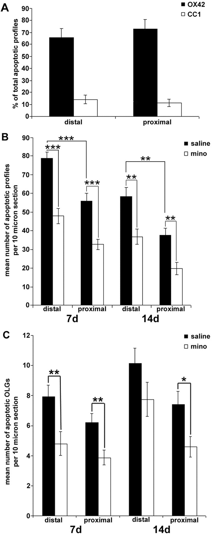
Figure 2.

Minocycline treatment inhibited dorsal column transection-induced glial cell death within the distal and proximal AST. A, Percentage of apoptotic microglia/macrophages (OX42-positive) or oligodendrocytes (CC1-positive) within the proximal and distal AST 7 d after the lesion (n = 6 per group). Microglia/macrophages are the main cell type to undergo apoptosis in this model. B, Quantification of the mean number of apoptotic profiles within the distal and proximal segments of the AST. Significantly fewer apoptotic profiles are located within the proximal versus distal segments of the AST. Minocycline (mino) treatment significantly reduced the mean number of apoptotic profiles per 10 μm section ± SEM within distal and proximal segments of the AST at both 7 and 14 d after injury compared with saline-treated controls. (*p < 0.05; **p < 0.01; ***p < 0.001; 7 d, n = 6 per group; 14 d, n = 4 or 5 per group). C, Apoptotic oligodendrocytes (CC1-positive), active caspase-3-positive profiles with a condensed nucleus, were significantly decreased in the minocycline-treated group compared with saline controls within the proximal AST at both 7 and 14 d after injury. However, only the proximal segment of the AST from minocycline-treated animals contained significantly fewer apoptotic oligodendrocytes at 14 d after injury. There was no difference between the mean number of apoptotic oligodendrocytes within the proximal versus distal segments of the AST at both 7 and 14 d after injury (data represent mean ± SEM; *p < 0.05; 7 d, n = 5 per group; 14 d, n = 4 or 5 per group).
