
Figure 3.
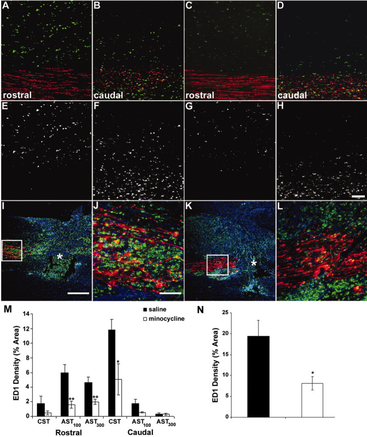
Minocycline treatment reduced ED1-positive (microglial/macrophage) density 7 d after injury. A-H, ED1-positive profiles (green) 3 mm rostral (A, C, E, G) and caudal (B, D, F, H) to injury. Minocycline significantly reduced ED1 density within both rostral AST (100 and 300 μm dorsal to CST; C, G, M) and within the caudal CST (D, H, M) compared with saline-treated animals (A, B, E, F, M). Triple immunofluorescence images of the lesion site from saline-treated (I, J) and minocycline-treated (K, L) animals are shown. The proximal CST is red; ED1-positive microglia/macrophages are green; and Hoechst 33258 is blue. Note the reduced ED1-positive signal in K compared with I. J, L, Higher magnification of the boxed area in I and K. Less ED1-positive signal is evident within the proximal CST of minocycline-treated animals compared with saline-treated animals. N, Quantification of the density of the ED1-positive signal within the proximal CST. Data represent mean percentage ± SEM (n = 4 or 5 per group). *p < 0.05; **p < 0.01. Scale bars: A-H, J, L, 100 μm; I, K, 500 μm.