
Figure 5.
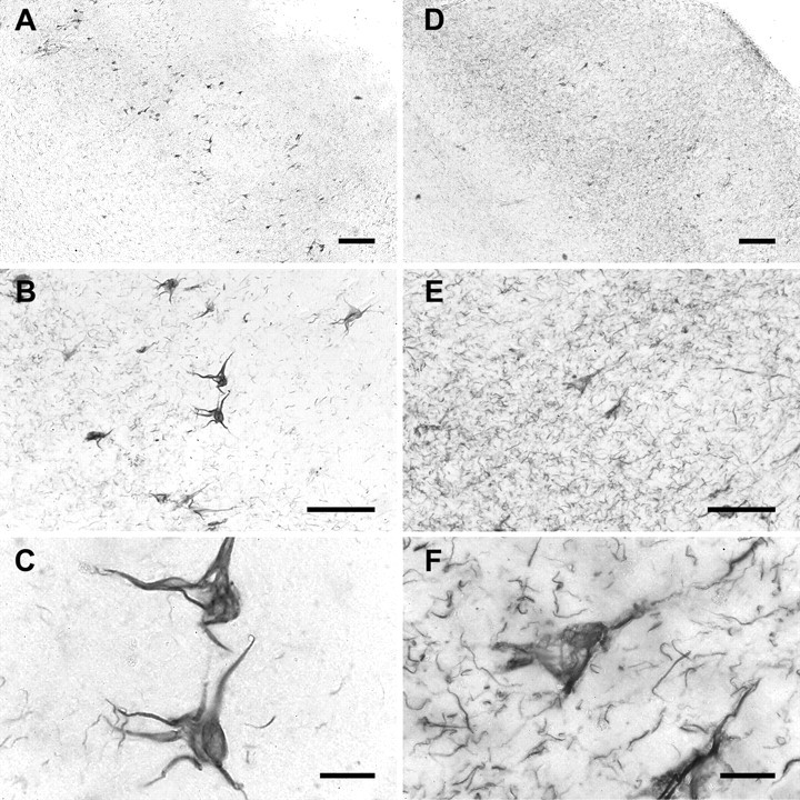
AD brain tissue contains tau phosphorylated at tyr18. Sections from the entorhinal cortex of AD brain were labeled with either 9G3 (left) or AT8 (right) followed by development using DAB and nickel ammonium sulfate (see Materials and Methods). Magnification in each left panel is identical to that of the adjacent right panel. Scale bars: A, D, 200 μm; B, E, 100 μm; C, F, 20 μm. Note that AT8 labeled neuropil threads and dystrophic neurites, whereas 9G3 did not.