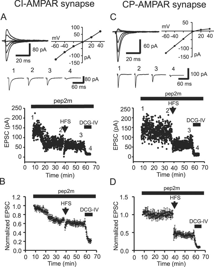
Figure 7.
Postsynaptic AMPAR trafficking contributes to expression of LTD at CI-AMPAR synapses. The NSF-AP2-inhibitory peptide pep2m (0.5 mm) was included in the recording pipettes for all experiments. A, CI-AMPAR synapse. Top, EPSCs evoked at holding potentials between -60 and +40 mV, recorded during the first 8 min after formation of whole-cell configuration (left) and the corresponding I-V relationship (right) from a CI-AMPAR synapse. dl-APV was included for the period of RI determination. Middle, Averaged current traces from 10 EPSCs taken at the time points indicated in the dot plot below. Bottom, Dot plot of EPSC amplitude indicating the time course of the experiment. The first 8 min after the formation of whole-cell recording were used to construct the I-V relationship and identify the Ca2+-permeable nature of the AMPAR-mediated EPSC. Inclusion of the NSF inhibitory peptide inhibited EPSCs at this CI-AMPAR synapse and occluded expression of LTD. DCG-IV was included in the extracellular solution at the end of the recording to confirm that EPSCs were mossy fiber in nature. B, Averaged data from seven CI-AMPAR synapses illustrate the reduction of EPSC amplitude and the concomitant occlusion of LTD. C, CP-AMPAR synapse. The I-V relationship and the time course of evoked EPSCs at a CP-AMPAR synapse are shown. Organization of data is identical to that shown in A and B. Inclusion of the NSF inhibitory peptide was without effect on both the EPSC amplitude and the ability to express LTD at CP-AMPAR synapses. D, Averaged data from five CP-AMPAR synapses.

