
Figure 1.
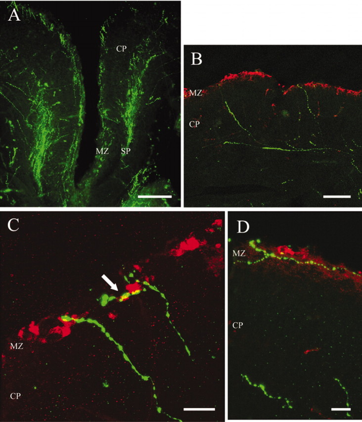
Serotonergic afferents (FITC, green) contact CR cells (Cy3, red) in the MZ of E17 embryos. A, Serotonergic fibers enter the developing cortex and form two bands, one in the marginal zone (MZ) and the other in the subplate (SP). B, Some serotonergic fibers in the SP traverse the cortical plate to reach the MZ. C, Confocal microphotograph of serotonergic fibers contacting CR cells in the MZ. The arrow indicates a point of contact confirmed by rotating the image in 3D. D, Horizontal serotonergic fibers contacting CR cells in the MZ. Scale bars: A, 200 μm; B, 100 μm; C, D, 20 μm.