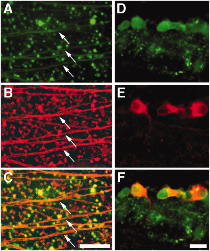
Figure 1.

AAV-mediated transfection of RGCs. A-C, Flat-mounted rat retina 3 weeks after intravitreal injection with AAV-NgRWT-IGFP, double-stained for GFP to detect transfected cells (A) and βIII tubulin, a selective marker for RGCs in the retina (B). Note staining of cell bodies and axon fascicles (arrows) throughout the retina. C, Merged image. D-F, Retinal cross section, double labeled as above. GFP-expressing cells (D) are located within the innermost retina and show positive staining for βIII tubulin (E). F, Merged image. Scale bars: A-C, 100 μm; D-F, 20 μm.
