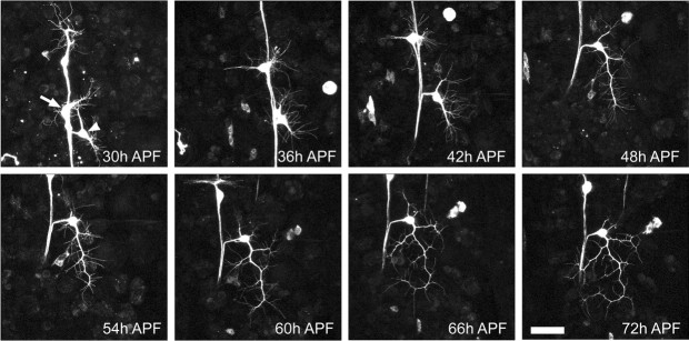
Figure 10.
Effect of 24 hr APF juvenile hormone mimic application on adult outgrowth of ddaE. Projections of multiphoton z-stacks of ddaE expressing the CD8::GFP reporter are shown. JHm was applied at 24 hr APF, and ddaE was imaged at 6 hr intervals over the period from 30 to 72 hr APF. At 30 hr APF, the larval dendrites have been pruned, and the cell body of ddaE has migrated dorsally with the stretch receptor dbd. A number of filopodia can be seen extending from the cell body. At 36 hr APF, the cell body has numerous filopodia extending from short primary dendrites. Between 42 and 60 hr APF, the arbor undergoes a gradual increase in complexity and size. Between 66 and 72 hr APF, the arbor reaches the edge of its normal footprint. Arrow denotes dbd. Arrowhead denotes ddaE. Scale bar, 40 μm.

