Figure 3.

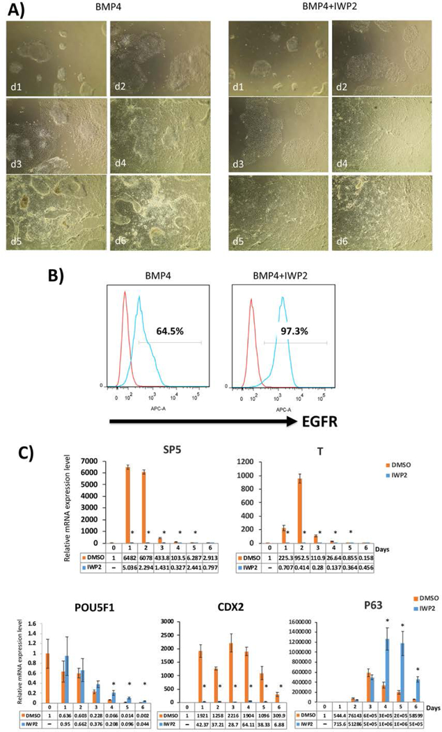
Comparison of trophoblast differentiation using BMP4, with and without IWP2, starting with E8-adapted WA09/H9 hESCs.
A) Morphology of the cells in IWP2 shows more uniform flattening starting from one day post-BMP4 treatment, in comparison to BMP4 alone. This morphological difference was consistent through all 6 days of the first step differentiation.
B) Expression of CTB marker, EGFR, as measured by flow cytometry on day 6 following BMP4 treatment. The addition of IWP2 leads to a higher number of EGFR+ cells.
C) qPCR for lineage-specific marker expression during the first-step differentiation with and without IWP2. Compared to the DMSO vehicle control, marker of WNT signaling, SP5, is suppressed in the presence of IWP2, as expected. T/Brachury, an early marker of mesoderm induction, is also suppressed. At the same time, CTB markers (p63 and CDX2) are significantly increased both with and without IWP2, although the fold change for CDX2 (also a marker of mesoderm) is lower in the presence of IWP2. Pluripotency marker, POU5F1, is appropriately decreased during differentiation. Data are normalized to 18S and expressed as fold-change above day 0 (undifferentiated state). *indicates P<0.05 compared to DMSO control at the same time-point.
