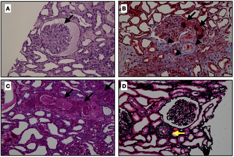
Figure 2.
Classic renal pathology in scleroderma renal crisis. (A) The glomerulus demonstrates the global collapse of the glomerular capillary with bloodless appearance, finding similar to ischemic changes. There is no crescent or proliferative lesion (Hematoxylin and eosin stain, x400). (B) This figure shows extensive fibrinoid necrosis and fibrin thrombi in afferent arteriole and glomerulus (arrows), and mucoid intimal edema of small arteries (arrow heads) (Masson’s trichrome, original magnification x400). (C) Obliteration of arterioles and interlobular arteries (arrows) with thrombi (Periodic acid-Schiff stain, original magnification x400). (D) Intimal concentric lamination (arrow) or onion skin appearance of arteriole (Methenamine silver stain, original magnification x400).

