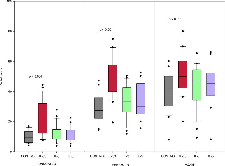
Fig 1. Eosinophil adhesion with IL-33.
Eosinophil adhesion determined by EPX release from adherent cells cultured for 30min with cytokine stimulation, cytokine concentration 1ng/mL. Values shown are a percentage of optical density (OD) of wells containing 10x10e4 lysed eosinophils (n = 22). Experiments from different blood donors with the exception of 3 donors that were repeated twice in which case, results were averaged from the 2 different experiments using their cells. Lines connect comparison groups with p-value denoting significant difference in pair-wise comparisons.

