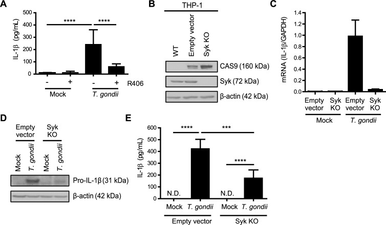
Fig 4. Syk contributes to IL-1β production in T. gondii-infected monocytic THP-1 cells.
(A) THP-1 cells were pretreated with 2 μM R406 or vehicle control for 40 min and then mock treated or infected with T. gondii for 18 h, and the levels of IL-1β in the supernatant were measured by ELISA. (B) Lysates from wild-type (parental) THP-1 cells, control Empty Vector THP-1 cells, and Syk KO THP-1 cells were blotted with antibodies to visualize Cas9, Syk, and β-actin. (C) Empty Vector THP-1 cells and Syk KO THP-1 cells were mock treated or infected with T. gondii and qPCR was performed with primers specific for IL-1β. Transcript levels relative to those of GAPDH are graphed. (D and E) Empty Vector or Syk KO THP-1 cells were mock treated or infected with T. gondii, and pro-IL-1β and β-actin in the cell lysate were visualized by Western blotting (D) or the levels of IL-1β in the supernatant were measured by ELISA (E). In (A) and (E), combined data from 4 experiments are shown. Representative Western blots (B and D) and qPCR (C) from 4 experiments are shown. Values are expressed as the mean ± SD, ***P<0.001, ****P<0.0001 (one-way ANOVA followed by a Tukey post-test in A and E).

