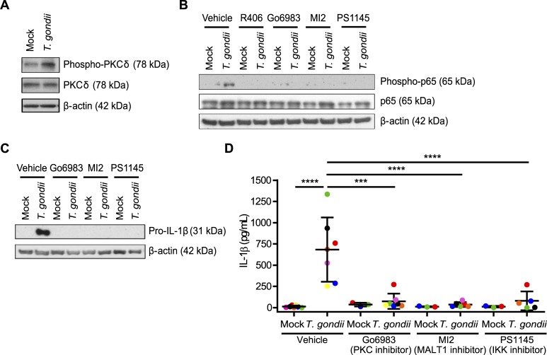
Fig 5. The Syk-PKCδ-CARD9/MALT-1-NF-κB pathway is activated in T. gondii-infected human monocytes.
(A) Primary monocytes were mock treated or infected with T. gondii and the lysates were blotted for total and phospho-PKCδ and for β-actin. (B) Primary monocytes were pretreated with 2 μm R406, 300 nM Go6983, 3 μM MI2, 100 nM PS1145, or vehicle control for 40 min then mock treated or infected with T. gondii for 1 h. Total and phospho-p65 (Ser536) and β-actin in the cell lysate were visualized by Western blotting. (C) Pro-IL-1β and β-actin were visualized by Western blotting of lysates from primary monocytes that were mock treated or infected with T. gondii for 4 hr in the presence of the vehicle control or the indicated inhibitors. (D) The levels of IL-1β in the supernatant of mock or T. gondii-infected primary monocytes in the presence or absence of the indicated inhibitors were measured by ELISA. Data in (D) are combined results of 3–7 experiments with independent donors. Representative Western blots from 3 (A and C) and 4 (B) experiments are shown. Values are expressed as the mean ± SD, ***P<0.001, ****P<0.0001 (one-way ANOVA followed by a Tukey post-test).

