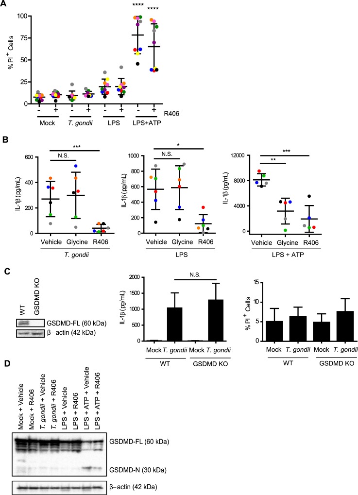
Fig 7. T. gondii-induced IL-1β release is independent of cell death and gasdermin D (GSDMD).
(A) Primary human monocytes were pre-treated with 2 μM of the Syk inhibitor R406 or vehicle control (DMSO) for 40 min, mock treated, infected with T. gondii, stimulated with LPS (100 ng/ml), or stimulated with LPS (100 ng/ml) plus ATP (5 mM) for 4 h (ATP was added for the last 30 minutes of treatment). Cell viability by PI staining was then measured by flow cytometry. (B) Primary monocytes were pretreated with 5 mM glycine, 2 μM R406, or vehicle control for 40 min. Cells were then infected with T. gondii, stimulated with LPS alone or LPS+ATP for 4 h, and the levels of IL-1β in the supernatant were measured by ELISA. (C) Lysates from wild type and GSDMD knockout (KO) THP-1 cells were blotted with anti-GSDMD or anti-β-actin antibodies. The WT or GSDMD KO cells were mock treated or infected with T. gondii for 18 h. The levels of IL-1β in the supernatant were measured by ELISA, and cell viability by PI staining was measured by flow cytometry. (D) GSDMD-FL (full-length) and GSDMD-N (N-terminal) protein in the cell lysate were visualized by Western blotting. Data in (A) and (B) reflect combined results of 9 and 5 experiments with independent donors, respectively. For (C), combined data from 6 and 13 experiments is shown for the IL-1β release ELISA and PI staining experiments respectively. In (D) a representative Western blot from 4 experiments is shown. Values are expressed as the mean ± SD, *P<0.05, **P<0.01, ***P<0.001, ****P<0.0001 (one-way ANOVA followed by a Tukey post-test in A, B and C). In (A), the LPS+ATP treated conditions contained a significantly higher percentage of PI+ cells than all of the first six conditions, which were not significantly different from each other.

