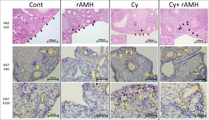
Fig. 3.
Histological evidence of follicle activation and rAMH protection in an in vivo mouse model of Cy treatment. Representative H&E images of ovaries from each of the four treatment groups indicating the changes in primordial follicle (black arrows) and primary follicle (red arrows) populations after treatment. Magnification of boxed areas is shown in the inset pictures. Immunohistochemical staining for Ki67 on ovaries removed 2 days after Cy treatment from mice from each treatment group. Yellow arrows indicate follicles containing granulosa cells which stained positive for Ki67. Sections from a minimum of four animals in each group were stained

