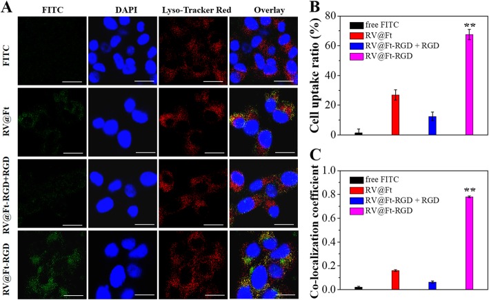
Fig. 4.
a Fluorescence images of A549 cells after incubation with free FITC and FITC labeled RV@Ft, RV@Ft-RGD + RGD, and RV@Ft-RGD, respectively. Scale bar = 60 μm. b FCM measurement of cellular FITC fluorescence intensities in A549 cells after incubation with free FITC and FITC labeled RV@Ft, RV@Ft-RGD + RGD, and RV@Ft-RGD, respectively. c Co-localization coefficient of cellular fluorescence of FITC and Lyso Tracker Red. **P < 0.01, compared with other groups, respectively

