
Figure 3.
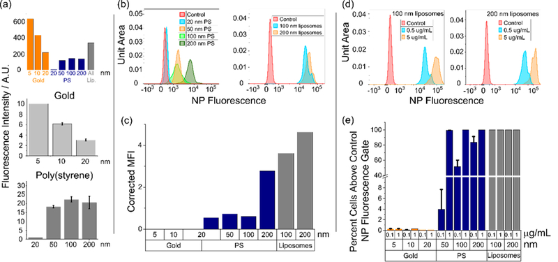
Neutrophil internalization of nanomaterials is size-dependent. All particle incubation times are 3 h unless otherwise noted. (a) Theoretical expected relative fluorescence intensities for different particle formulations as they would be observed by a flow cytometer, calculated for fixed particle concentrations with known detector gains, fluorophore properties, and cytometer excitation/emission wavelengths (top). Comparison of actual fluorescence emission intensities observed at 1.0 μg/mL for gold (middle) and poly(styrene) (bottom) particles on a fluorescence plate reader. (b) NP fluorescence intensity distribution histograms for poly(styrene) and liposomes taken up by nonapoptotic, nonactivated neutrophils as a function of particle size. PS particles incubated at 1 μg/mL. Liposomes incubated at 0.5 μg/mL. (c) Mean NP fluorescence intensities as observed by the flow cytometer for nonapoptotic, nonactivated neutrophils. Particle concentrations were 1.0 μg/mL. Fluorescence intensities as observed by the cytometer were adjusted by subtracting the untreated control fluorescence and applying the appropriate correction factor calculated in Figure 3a. (d) Dose response of NP fluorescence intensities for liposomes in nonapoptotic, nonactivated neutrophils. (e) Dose response for gold, PS, and liposomal formulations in nonapoptotic, nonactivated neutrophils. Percent cells with fluorescence exceeding the gate (maximum value of the control) calculated according to the gating shown in Figures 2a–c. Particle incubation time was 12 h.