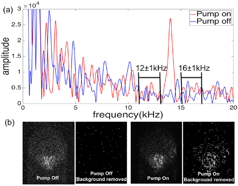
Fig. 2.
(a) Frequency domain signal from one pixel in the MB filled capillary tube with and without the pump on. The relevant aspects of the background subtraction algorithm are labeled where the background is assumed to vary linearly over a narrow frequency range. (b) comparison of PPOCT images with background subtraction demonstrating the complete removal of the of noise background.

