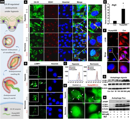
Fig. 3. Nanofibers promote CA IX–regulated endocytosis under hypoxia.

(AW) Artwork here illuminates the whole process of CA IX-regulated endocytosis for nanofibers under hypoxia. (A) CA IX–regulated endocytosis of N-pepABS nanofibers has appeared under hypoxia after 24-hour treatment of 500 μM N-pepABS (+) or medium control (−). Scale bar, 20 μm. We further observe subsequent (B) endolysosomal swelling and (C and D) intracellular acid vesicles injuries after 48-hour treatment of 500 μM N-pepABS under hypoxia. Scale bars, 20 μm. Then, blockage of protective autophagy has been detected after 48-hour treatment of 500 μM N-pepABS. (E) Ratio of mRNA levels of Atg5/GADPH, with the bar image represented as means ± SD, while *P < 0.05 was thought as significant difference. (F) Fluorescence images of autophagosome accumulation in the cytoplasm of hypoxic cancer cells. Scale bars, 20 μm. LC3B, light chain 3B. (G) Western blot assays of autophagy-related signals in MDA-MB-231 cells and their (H) autophagy flux study with 1-hour pretreatment of 10 nM bafilomycin A1 (Baf). cPARP, cleaved poly(adenosine diphosphate–ribose) polymerase.
