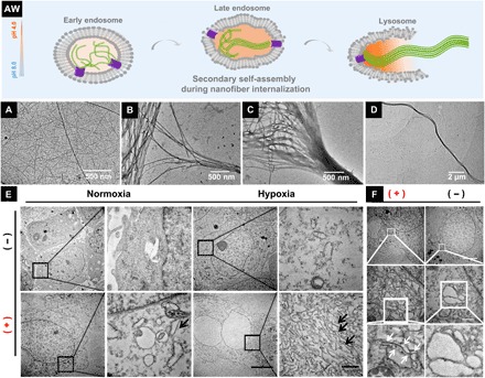
Fig. 4. CA IX–induced nanofiber internalizations in hypoxic cancer cells.

(AW) Hypothesis of structure upgrades during CA IX-induced nanofiber internalization. TEM images of nanofibers formed by 0.75 wt % of N-pepABS at (A) pH 6.5 or (B to D) pH 5.5. TEM images of nanofibers (E) dissociatively appearing in the cytoplasm (black arrows) and (F) destroying monolayer vesicles (white arrows) of hypoxic MDA-MB-231 cells, after 48-hour treatment of 500 mM N-pepABS. Scale bars, 5 μm (E) and 500 nm (F).
