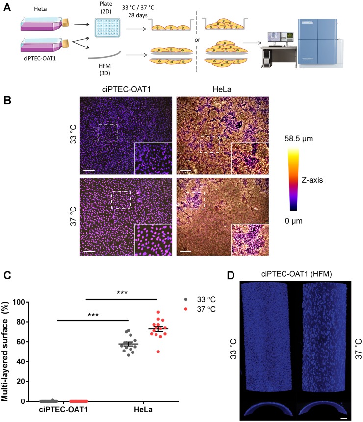
Figure 2. Contact inhibition in ciPTEC-OAT1.
(A) Schematic diagram of focus formation assay. CiPTEC-OAT1 were cultured in 2D (96-well microplate) and 3D (hollow fiber membranes; HFM) for 28 days at 33° C and 37° C. HeLa cells were cultured in 2D in same conditions. Foci (multi-layered growth) formation was detected by nuclear staining and confocal imaging and (B) representative depth-coded images of nuclei-stained ciPTEC-OAT1 and HeLa cells after 28 days of culture at both permissive and non-permissive temperature are shown. Scale bars denote 200 μm in the original image and 100 μm in the zoom-in. (C) Quantification of the surface area covered by multi-layered proliferation). ND = not detected. (D) Representative confocal images of nuclei stained ciPTEC-OAT1 cultured on double-coated HFM at 33° C and 37° C, x-y confocal planes on the upper part and y-z confocal planes on the bottom part. Images taken with 10× magnification. Scale bar: 50 μm. Values are expressed as the mean ± SEM of three independent experiments performed in triplicate. *** p < 0.001 (unpaired two-tailed Student’s t-test).

